Coronary AngioTAC
What does Coronary Computed Axial Tomography (Coronary Angio CT) consist of?
In the last decade, coronary CT has had an important role in the non-invasive diagnostics of various pathologies associated with the risk of sudden death, especially thanks to the substantial reduction in exposure to ionizing radiation obtained through the use of low voltage tubes and innovative image reconstruction techniques. For example, the PROTECTION VI multicenter registry showed that from 2007 to 2017, the average exposure to ionizing radiation for an examination of coronary angiography by CT decreased by 78%, from 885 mGy * cm to 195 mGy * cm.
Similar to Nuclear Magnetic Resonance, Cardiac Computed Tomography allows an accurate quantification of volumes, ejection fraction and biventricular mass. In addition, it enables the evaluation of the origin of the coronary arteries, and above all, the coronary circulation by quantifying the extent of any luminal atherosclerotic obstructions, using the Coronary Angiography CT (CCTA) technique, and the degree of calcification, by means of the technique of Calcium Scoring TC (CCS).
The technique of calcium scoring through validated scores, such as the Agatston score, allows a quantification of coronary calcifications which were found to be a valid element in predicting the risk of myocardial infarction and sudden death independently of traditional risk factors.
What are the main applications of Coronary Computed Tomography (Coronary CT)?
The main applications of cardiac CT are:
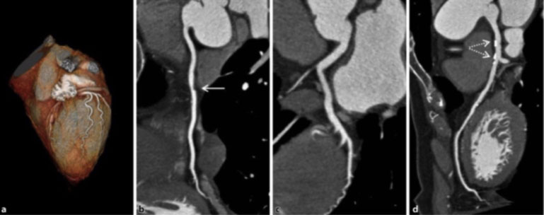
1 – Coronary artery atherosclerotic disease associated with stable angina: In the patient with stable angina and low-intermediate risk of obstructive coronary artery disease, the CCTA 64 slices has a sensitivity of 98%, a specificity of 90% in detecting a significant obstructive coronary artery disease and a negative predictive value greater than 95% (1). This means that, in these patients, a clinically relevant coronary obstruction can reasonably be excluded in the presence of negative findings.
| For this reason, the 2013 European Guidelines place the coronary assessment by CCTA in the low-intermediate risk patient on the same level as the stress test (Figure 1). |
Adding to this the possibility of studying coronary perfusion through the administration of vasodilating agents and iodinated contrast medium, it is possible to obtain functional, as well as anatomical, information related to the coronary obstructive plaque and the myocardium dependent on it for perfusion (2). However, this technique is rarely used to date, especially in consideration of the fact that it is possible to evaluate myocardial perfusion in an equally non-invasive way and without the administration of ionizing radiation using specific protocols of cardiac MRI.
2 – Patient with acute chest pain: Several randomized trials worldwide have assessed the role of CCTA in assessing patients with acute chest pain. One of the most important is the CATCH trial, which assessed the use of CCTA in the patient with acute chest pain with presentation in the emergency / urgency department with normal cardiac enzymes and normal or non-specific ECG findings. In these patients, the CCTA has proven to be able to safely rule out an acute coronary syndrome as demonstrated by an 18.7-month follow-up free from cardiovascular events (3). Furthermore, it should not be forgotten that in the setting of the patient with acute chest pain, computed tomography can be used for the differential diagnosis with other syndromes, such as acute aortic dissection and pulmonary embolism. In fact, it is possible to use a triple rule-out protocol which, through specific sequences, allows to study at the same time the presence of coronary atherosclerotic disease, aortic dissection and pulmonary embolism. Currently this protocol is applied only to 0.5% of the cases in question, most likely due to the higher doses of contraction medium required, the long apnea times and the increase in ionizing radiation administered.
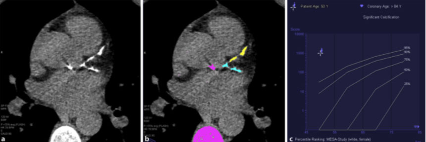
3- Stratification of asymptomatic patients with intermediate-high cardiovascular risk profile: Calcium Scoring TC is a technique that by quantifying the presence of coronary calcifications allows to stratify patients with an intermediate-high cardiovascular risk profile, but still asymptomatic, and thus guide the intensity of preventive medical treatments (4). In fact, from a clinical point of view, the stratification of cardiovascular risk is primarily based on the analysis of traditional risk factors, such as diabetes, smoking, hypertension, dyslipidemia and the family history of atherosclerotic coronary artery disease using score systems, such as the EUROPEAN score, which, taking into account age and gender, are able to define the risk at 10 years.
The purpose of the definition of CCS in asymptomatic intermediate-high risk patients is therefore to add an incremental risk to the analysis of traditional cardiovascular risk factors: patients who would benefit from aggressive treatment of these risk factors, in particular cholesterol and blood pressure, as well as promoting a change in lifestyle (Figure 2).
CCS is therefore a strong prognostic factor and it was recently included in the 2018 American guidelines for the management of cholesterol levels (5). Low-risk patients do not benefit from this type of analysis, and high-risk or symptomatic patients obtain greater benefits from performing coronary angiography.
4 – Abnormalities of coronary artery origin: CCTA is the method of choice for the study of suspected anomalies of coronary origin. In particular, it allows the evaluation of the coronary anatomy and the exact course of the vessel, a particularly important element in cases of inter-arterial passage, i.e. between the trunk of the pulmonary artery and the ascending aorta. This course is associated with the risk of myocardial ischemia and sudden death (6).
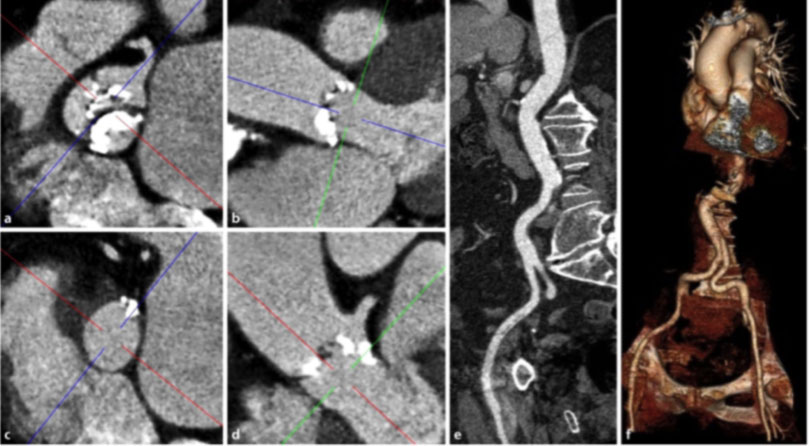
5- Preoperative assessment in cases of percutaneous aortic valve replacement (TAVI): The aortic angio CT is the method of choice in the case of preventive assessment of percutaneous aortic valve implantation (TAVI), as it allows a three-dimensional evaluation of the aortic root, aortic annulus and ileofemoral vascular accesses (7, Figure 3 ).
6- Congenital heart malformations of children and adults: Cardiac CT obtains precise three-dimensional reconstructions of congenital heart disease. The need to administer a low dose of ionizing radiation, however, means that, currently, echocardiography and magnetic resonance imaging are still the preferred imaging methods in this regard. To date, it plays a fundamental role, especially in those patients in whom the presence of contraindications exist for echocardiography and cardiac magnetic resonance imaging (8).
What are the main advantages of coronary CT?
The main advantage of cardiac CT is that it is a non-invasive examination, which allows to accurately evaluate cardiovascular risk, calcium accumulation (CCS) and coronary anatomy, examining the state of the vessel wall, in addition to the characteristics of their lume (the internal space), giving a semi-quantitative assessment of the degree of stenosis (narrowing).
What are the risks of coronary CT?
Coronary CT is not painful or invasive. A potential problem is an allergy related to the contrast medium, for which a specific preparation must be carried out. Any known allergies must be reported at the time of booking.
What is the preparation for coronary CT?
The patient must completely fast for at least 6 hours before the exam. It is not necessary to stop taking medications in use (e.g. anti-hypertensives), which must be taken only with a little water.
On the day of the examination, the patient must bring the previous radiological examinations (X-rays, CT, Resonances, Ultrasounds, Visits, etc.) with them, even if performed elsewhere and have the result of the blood chemistry tests: Urea and Creatinine.
The patient must have the prescription of the attending physician (in the case of an examination with the NHS) or a simple specialist prescription (in the case of a private examination)
The duration of the exam is approximately 30 minutes.
The patient must be accompanied by a trusted person, since during the examination drugs are administered that can influence psychophysical abilities. Thus, it is advisable not to be alone and not to drive immediately after the examination.
In the case of patients with very frequent arrhythmias or with rapid atrial fibrillation, an optimization of the therapy may be necessary to control the heart rate, to be carried out according to the advice of the treating cardiologist.
If you need to book a Coronary AngioTAC -> Information request and / or exam


Bibliography:
-
- Head-to-head comparison of prospectively triggered vs retrospectively gated coronary computed tomography angiography: Meta- analysis of diagnostic accuracy, image quality, and radiation dose. Menke J, Unterberg-Buchwald C, Staab W et al (2013). Am Heart J 165:154–163 (e153) Varga-Szemes A, Meinel FG, De Cecco CN et al (2015) CT myocardial perfusion imaging. Ajr Am J Roentgenol 204:487–497.
- CT myocardial perfusion imaging. Varga-Szemes A, Meinel FG, De Cecco CN et al (2015) Ajr Am J Roentgenol 204:487–497
- Long- term clinical impact of coronary CT angiography in patients with recent acute-onset chest pain: the randomized controlled CATCH trial. Linde JJ, Hove JD, Sørgaard M et al (2015). JACC Cardiovasc Imaging 8:1404–1413
- Coronary calcium score and cardiovascular risk. Greenland P, Blaha MJ, Budoff MJ et al (2018). J Am Coll Cardiol 72:434–447.
- Guideline on the management of blood cholesterol: a report of the American college of cardiology/American heart association task force on clinical practice guidelines. Grundy SM, Stone NJ, Bailey AL et al (2018) AHA/ACC/AACVPR/AAPA/ABC/ACPM/ADA/AGS/ APhA/ASPC/NLA/PCNA. J Am Coll Cardiol pii:S0735-1097(18)39034-X.
- Anomalous coronary arteries that need interven- tion: review of pre- and postoperative imaging appearances. Agarwal PP, Dennie C, Pena E et al (2017) Radiographics 37:740–757.
- Computed tomography imaging in the context of transcatheter aortic valve implantation (TAVI)/ Transcatheter aortic valve replacement (TAVR): an expert consensus document of the society of cardiovascular computed Tomography. Blanke P, Weir-Mccall JR, Achenbach S et al (2019). Jacc Cardiovasc Imaging 12:1–24.
- ECG- synchronized CT angiography in 324 consecutive pediatric patients: Spectrum of indications and trends in radiation dose. Meinel FG, Henzler T, Schoepf UJ et al (2015). Pediatr Cardiol 36:569–578.
