Electrophysiological Study
Indice dell'articolo
FOR WHAT IS THE ENDOCAVITARY ELECTROPHYSIOLOGICAL STUDY (EES)?
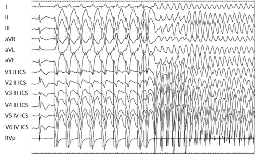
The endocavitary electrophysiological study (also known by the acronym EES) is an invasive diagnostic test that evaluates the electrical properties of the heart and its susceptibility to developing different types of cardiac arrhythmias. The purpose of the EES is to identify the nature of the arrhythmic disorder and its place of origin in the heart. This may require the investigated arrhythmia to be induced by electrical impulses delivered from the catheter tip during the examination. In most cases, the arrhythmia induced during the electrophysiological study is interrupted using the same impulses that generated it. Occasionally, an electric shock in short narcosis (without intubation) may be needed to end the arrhythmia. The identification of the arrhythmia by this study will enable the arrhythmologist to decide the most appropriate therapy for its abolition and / or prevention. In some cases, it will be possible to cure the arrhythmia definitively by using one or more (painless) supplies of radiofrequency energy that represents the transcutaneous ablation procedure.
HOW IS THE ELECTROPHYSIOLOGICAL STUDY PERFORMED?
The electrophysiological study takes place in the electrophysiological room or laboratory, after a short hospitalization. After performing local anesthesia, two to five thin catheters are inserted in the right heart (electrical cables covered with a plastic sheath) which are made to proceed under a permanent radiological control (X-rays). The pathways necessary to reach these heart catheters include the right femoral veins (at the level of the groin), the left subclavian vein (at shoulder level) and rarely the left or right jugular vein (at neck level). An electrical signal is then recorded through the tip of each catheter that originates from the cardiac cavity in which it is located.
WHEN IS THE ELECTROPHYSIOLOGICAL STUDY PERFORMED?
The electrophysiological study can be performed in a patient at risk of cardiac arrhythmias with different clinical indications:
* DIAGNOSTICS: when the electrophysiological study can be used for the purpose of performing an arrhythmogenic diagnosis that cannot be performed using non-invasive methods (for example, dysfunctions of the sinus node that can occur with dizziness or syncope, the atrioventricular blocks that are indicated for a definitive pacemaker implant) , tachycardias, ventricular extrasystoles, etc.);
* THERAPEUTIC: when the electrophysiological study is performed for the purpose of establishing adequate therapy, whether pharmacological, surgical or performed during the electrophysiological study itself (for example the ablation of an anomalous pathway or tachycardia);
* PROGNOSTICS: when the electrophysiological study aims to establish the risk of sudden death during ventricular arrhythmias.

WHAT ARE THE RISKS OF ELECTROPHYSIOLOGICAL STUDY?
The endocavitary electrophysiological study and the possible radiofrequency catheter ablation have, like all invasive procedures, a risk, even if minimal, of complications. The most frequent complications are the local ones that include a small hematoma at the catheter introduction site (incidence 7%), while, much more rare (<0.5% overall) are the lesions of the blood vessels or nerves that run near the vessels. Injuries to the vessels near the heart or in the heart itself occur at an extremely low frequency (1.2 cases per 1000 procedures). Usually, complications are transient (mild hematoma with self-absorption, transient chest pain) or correctable.
In summary, the risk associated with the electrophysiological study is very low and the advantage derived from its use for the patient is considerable.
