Late Potentials
What is the high resolution ECG for the study of late potentials?
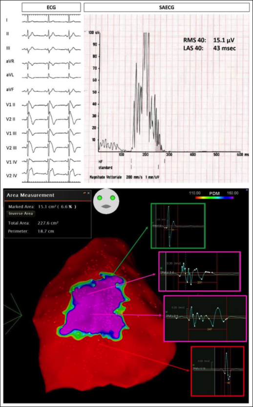
The high resolution ECG with recording (called SAECG, or “signal averaging ECG”) consists of a particular electrocardiographic recording that enables the identification of low voltage signals in the terminal part of the QRS complex of the electrocardiogram. This method is used in order to detect abnormal electrical activity in the heart muscle, especially at the ventricles, known as Late Ventricular Potentials (LVP, or “Ventricular Late Potentials”).
What are the Ventricular Late Potentials?
Late ventricular potentials are high frequency and low amplitude electrical signals, generally invisible on the standard electrocardiogram, but which can be detected thanks to electrocardiographic signal amplification techniques. The presence of late potentials on the path indicates the existence, in the ventricles, of an area in which electrical conduction is altered. The presence of late potentials identifies patients susceptible to develop serious ventricular rhythm disorders.
How are late potentials recorded and measured?
The search for LVPs is carried out with a special electrocardiograph that records about 1600 consecutive heart beats (for about 30 minutes) and analyzes cardiac activity with particular amplifications and filters in search of late potentials.
In our center the SAECG is carried out with ELI 350 SAECG System devices (Mortara Instruments Europe, Italy). The analysis is based on the acquisition of high resolution signals (1000 Hz) of the three orthogonal derivations (x, y, z) and the calculation of their average vector. The signals are then filtered with special filters to reduce noise (Band-pass filter frequency at 40–250 Hz bilateral filtering).
The SAECG algorithm automatically measures the following parameters:
- duration filter (f-QRSd);
- voltage of the quadratic root medium (root mean square voltage) of 40 ms QRS terminals filtered (RMS40)
- duration of low amplitude signals (<40 mV) in the terminal component of the filtered QRS (LAS40).
LVPs are considered present (pathological) when both of these conditions are present:
>> RMS40 < 20 mV & LAS40 > 38 ms
Under what conditions are late potentials sought after?
LVPs are sought in patients at risk for malignant ventricular arrhythmias. The positivity of late potentials indicates a greater degree of risk and can predict the inducibility of ventricular fibrillation. LVPs can be positive in particularly the following diseases:
- Brugada syndrome
- Arrhythmogenic Cardiomyopathy (or Dysplasia) of the right ventricle
- Idiopathic dilated cardiomyopathy
- Heart attack
- Congenital heart disease, for example Tetralogy of Fallot
Can the research for late potentials be useful in Brugada syndrome?
Our center recently published a study on LVP research in Brugada Syndrome. The results of this study show that LVPs were highly correlated with the arrhythmogenic substrate of Brugada syndrome, which is identified through epicardial mapping.
Therefore, the LVPs registered with a non-invasive method could be a useful tool to identify subjects at risk of malignant ventricular arrhythmias, as an expression of an altered electrical activity at the epicardial level.

Therefore, VPTs discovered with a non-invasive method could be a useful tool to identify subjects at risk of malignant ventricular arrhythmias, as an expression of an altered electrical activity at the epicardial level.
Does the exam to detect late potentials carry some risk?
The examination, like standard electrocardiography, is painless, free of side effects and does not require hospitalization. The duration of the exam is approximately 30 minutes. The exam does not require any preparation. The only collaboration required of the patient is to remain still and calm during the acquisition of the ECG trace.
If you need to book a Late Potential -> Request information and / or Exam
Follow the link for our recent article on late potentials in Brugada syndrome 2019 Europace Ciconte LP Brugada
If you need to book a Late Potentials click here -> Request for information
